Think about the last time you used a camera with actual film. It feels like a lifetime ago, right? Now, you pull out your smartphone, snap a picture, and see it instantly. That’s the exact same kind of leap dentistry has made with digital dental imaging.
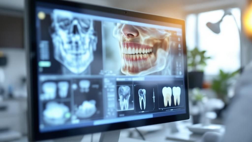
This technology uses tiny electronic sensors to capture incredibly clear pictures of your teeth, jaw, and gums, displaying them on a monitor in real-time for both you and your dentist to review together.
Understanding Modern Dental Imaging
Digital dental imaging is truly the backbone of modern, patient-first dental care. It’s a complete departure from the old, slow, and chemical-heavy process of traditional film X-rays. For dentists, it’s a powerful tool that allows us to diagnose problems faster, with greater accuracy, and with much less radiation exposure for you.
But this tech does more than just take pictures—it creates a detailed digital map of your entire oral health. These instant images become a permanent, secure part of your patient record. This makes it easy to track even subtle changes over time and to share information seamlessly with specialists if needed. The level of clarity and detail is what allows us to provide top-tier care.
The Shift to a Digital Standard
The move away from film reflects a huge step forward for the entire dental field. Instead of making you wait while film develops in a darkroom, we can now view, zoom in, and even enhance images right on the screen. This immediate feedback helps us be better diagnosticians and makes the whole appointment more comfortable and educational for you.
This shift empowers patients by allowing them to see exactly what their dentist sees. When you can view a high-resolution image of your own tooth on a screen, it becomes much easier to understand your diagnosis and treatment options.
To put the differences in perspective, let's compare the old way with the new.
Traditional Film vs Digital Imaging at a Glance
This table breaks down the key advantages of digital imaging over the traditional film X-rays you might remember.
| Feature | Traditional Film X-Rays | Digital Dental Imaging |
|---|---|---|
| Speed | Slow; requires chemical development (5-10 minutes). | Instant; images appear on-screen in seconds. |
| Image Quality | Fixed quality; cannot be adjusted or enhanced. | High resolution; can be zoomed, sharpened, and color-adjusted. |
| Radiation | Higher radiation dose. | Up to 90% less radiation exposure. |
| Patient Comfort | Bulky, sharp-edged film packets. | Smaller, rounded, and more comfortable sensors. |
| Diagnosis | Relies on a single, static image. | Allows for immediate, dynamic review and collaboration. |
| Storage & Sharing | Physical films are bulky and hard to share. | Digital files are stored securely and shared instantly. |
| Environment | Uses hazardous chemicals for developing. | Eco-friendly; no chemicals required. |
As you can see, the benefits are clear. Digital imaging isn't just a minor upgrade; it's a fundamental improvement in how we deliver care.
A Growing Field of Technology
The adoption of digital imaging in dentistry is already widespread and it’s not slowing down. The global dental imaging market is projected to grow from USD 3.26 billion in 2025 to nearly USD 4.69 billion by 2030. This growth is fueled by constant innovation, bringing more precise tools and better workflows to dental professionals.
This evolution covers everything from standard 2D bitewing X-rays to incredibly detailed 3D scans. A major part of this advancement is CBCT, or Cone Beam Computed Tomography. If you want to understand the cutting edge of modern dentistry, learning What is a CBCT? is a great place to start, as it provides the powerful three-dimensional views we use for complex procedures like dental implants and root canals.
Exploring Different Types of Dental Imaging
Think of digital dental imaging less as a single tool and more like a sophisticated toolkit. Just like a mechanic reaches for a specific wrench to fit a certain bolt, your dentist chooses the right imaging technology for your unique diagnostic needs. The mission is always the same: get the clearest possible picture with the least hassle and most comfort for you.
These technologies generally fall into two camps, defined by where the sensor is placed: inside or outside your mouth. This simple difference allows for a huge range of views, from a super-detailed close-up of one tooth to a sweeping overview of your entire jaw and facial bones. Knowing the difference helps take the mystery out of what’s happening during your appointment.
Intraoral Imaging: The Close-Up View
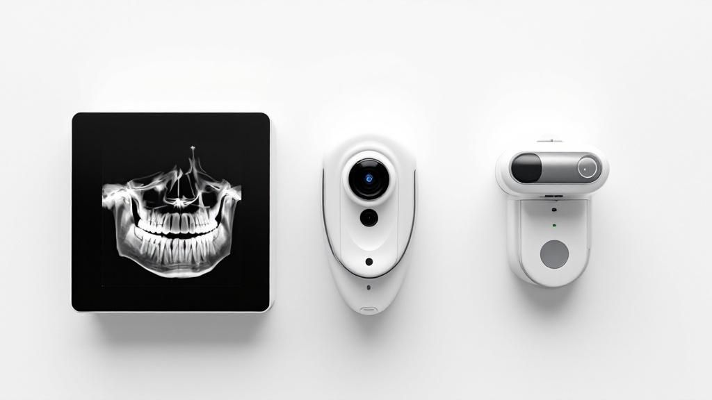
Intraoral imaging is what most people picture when they think of dental X-rays. It’s the workhorse of routine check-ups and targeted problem-solving. True to its name, a small, comfortable electronic sensor is placed inside your mouth to capture incredibly detailed images.
This is the dental equivalent of a high-powered magnifying glass. It’s perfect for any task that demands precision, such as:
- Finding Cavities: Spotting sneaky decay between teeth, where problems love to hide.
- Checking Tooth Roots: Making sure the root and the bone around it are healthy.
- Assessing Gum Health: Evaluating bone levels to screen for periodontal disease.
You’re probably familiar with bite-wing X-rays, which show the crowns of your top and bottom teeth in one go, and periapical X-rays, which give us the full picture of a tooth from the crown all the way to the tip of the root.
Extraoral Imaging: The Big Picture
Sometimes, we need to see the forest, not just the individual trees. That’s where extraoral imaging comes in. For these scans, the imaging machine stays outside your mouth, capturing a wide, comprehensive view of your jaw, teeth, and skull.
The classic example is a panoramic X-ray. You’ve likely had one of these—the machine rotates around your head to create a single, flat image of your entire mouth. It’s incredibly useful for:
- Planning orthodontic treatments like braces.
- Getting a good look at impacted wisdom teeth.
- Spotting issues with the jaw joint (TMJ) or cysts.
This type of imaging is less about the microscopic details and more about understanding the overall architecture and how all the different structures in your mouth and jaw relate to one another.
The global market for dental digital X-rays, covering both intraoral and extraoral systems, was valued at USD 3.45 billion in 2025. It’s expected to grow at 7.68% annually, hitting around USD 6.7 billion by 2034. This trend highlights the industry’s massive shift toward these faster, safer, and more precise technologies. You can discover more about this market growth and how it’s changing patient care for the better.
Cone Beam CT: The 3D GPS for Your Mouth
The most advanced tool in our digital imaging arsenal is Cone Beam Computed Tomography (CBCT). This technology is a true game-changer, taking us from flat, 2D pictures into the third dimension. A CBCT scanner rotates around your head, capturing hundreds of images that powerful software then stitches together into an interactive 3D model.
Imagine your dentist having a detailed, virtual map of your entire mouth and jaw that they can spin, slice, and zoom in on. This gives us an unbelievable level of detail, allowing us to see nerve pathways, bone density, and tooth orientation with absolute clarity. This kind of precision is crucial for complex procedures, including:
- Dental Implant Placement: Making sure the implant is positioned perfectly, avoiding any damage to nerves or sinuses.
- Complex Root Canals: Navigating tricky, curved, or hidden root canal systems.
- Surgical Planning: Creating a detailed anatomical roadmap to guide extractions or jaw surgery.
By giving us a true three-dimensional view, CBCT all but eliminates the guesswork that used to be part of complex dentistry, leading to far more predictable, safe, and successful results for patients.
How the Digital Imaging Workflow Works
So, what actually happens between the moment a sensor is placed in your mouth and the crystal-clear image popping up on your dentist's screen? It’s a remarkably fast and efficient process that has completely changed how we diagnose and plan treatments. The entire digital imaging workflow is a seamless, three-part journey: capturing the image, refining it, and using it to map out your care.
You can think of it like a high-end digital photography session. First, there's the photoshoot itself (capturing the image). Next comes the editing phase (enhancing it for clarity). Finally, the photo is used for its ultimate purpose (in our case, diagnosis and treatment). Each step is quick, digital, and perfectly connected to the next.
From Sensor to Screen: The Capture and Processing Phase
The workflow kicks off with image acquisition. A small, comfortable digital sensor is placed inside your mouth, and it captures the necessary X-ray data in just a few seconds. Unlike old-school film that had to be developed in a darkroom full of chemicals, this data is instantly sent straight to a computer.
Now for the magic: image processing. Specialized software immediately translates that raw data into a high-resolution image. This is where your dentist can really dig in, using powerful tools to analyze every detail. They can zoom in on a single tooth, adjust the brightness and contrast to spot subtle decay, or apply filters to highlight bone density. This is what turns a good image into an incredible diagnostic tool.
The ability to instantly manipulate and enhance a digital image is a core advantage. It allows dentists to identify potential issues that would be virtually invisible on a static, one-and-done film X-ray, leading to earlier and more accurate diagnoses.
This simplified process visualizes the journey from capture to diagnosis.
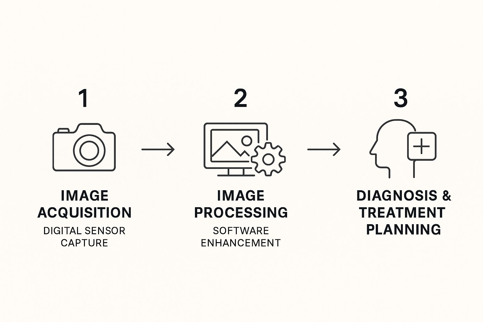
As the graphic shows, each step flows logically into the next, creating a streamlined and highly effective system for modern patient care.
Diagnosis, Storage, and Collaboration
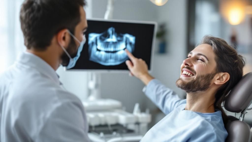
Once the image is ready, it's time for diagnosis and treatment planning. Your dentist can pull up the image on a large screen and walk you through exactly what they're seeing. They can point out areas of concern and clearly explain your treatment options. This shared view makes you an active, informed participant in your own healthcare journey.
Afterward, these digital files are stored securely in your electronic health record. This creates an invaluable timeline of your oral health, making it easy to track changes over the years. If a specialist's opinion is needed, the images can be shared instantly and securely, allowing for collaborative care without the usual delays.
Of course, a smooth workflow is about more than just capturing images; it’s about managing the data efficiently. Practices can seriously improve their operations when they automate data entry, which ensures patient records stay accurate and up-to-date. This frees up the team from administrative headaches so they can focus more on what matters—you.
The Payoff: A Better Experience for Patients and Practices Alike
Moving to digital dental imaging isn't just a simple tech upgrade—it fundamentally changes the dental experience for the better, both for you and for us. The advantages for patients are immediate and clear, starting with a massive improvement in safety. This modern approach cuts radiation exposure by as much as 90% compared to old-school film X-rays.
And let's be honest, no one will miss biting down on those sharp, uncomfortable film packets. Digital sensors are smaller, designed with rounded edges for comfort, and the images pop up on the screen instantly. No more waiting around for films to develop. It all adds up to less time in the chair and a much more pleasant visit.
Putting You in the Driver's Seat with Co-Diagnosis
One of the most powerful shifts with digital imaging is that you get to see exactly what your dentist sees. Instead of just hearing a diagnosis, you'll see the images on a large screen while your dentist walks you through every detail in real-time. This process, which we call co-diagnosis, transforms you from a passive patient into an active, informed partner in your own healthcare.
When you can clearly see a hairline fracture or the very beginning of a cavity, treatment recommendations just make sense. This visual proof empowers you to make decisions about your oral health with confidence.
This interactive, see-it-for-yourself approach is a cornerstone of modern dentistry. It builds a foundation of trust and makes sure you and your dental team are always on the same page, working toward the same health goals.
How It Elevates Care and Efficiency for Dental Practices
For a dental practice, the benefits are just as significant. It’s not just about fancy new tools; it’s about improving the quality of our care and how smoothly our office runs. The high-resolution, adjustable images lead to faster, more accurate diagnoses. We can spot problems far earlier, when they are smaller and much easier to treat.
Here’s a look at how it makes a difference in the day-to-day at a clinic like Beyond Dental Care:
- Faster Appointments: Capturing images instantly means no more waiting. This helps keep appointments running on schedule and shows we respect your time.
- Sharper Diagnostics: The ability to zoom in and adjust the contrast on an image helps us find tiny cracks or hidden infections that might have been missed on film. It’s what allows us to plan a dental implant with absolute precision for a perfect fit.
- A Simpler Workflow: Digital files are a breeze to store, organize, and share with specialists. This improves teamwork and coordinated care without the fuss of physical mail or lost records.
- A Safer Office: Getting rid of the harsh chemicals needed for film development makes the dental office a safer place for our team and is much better for the environment.
By weaving this technology into our practice, we can deliver a higher standard of care while creating a more streamlined, patient-first experience from the moment you walk in the door.
Connecting Imaging to the Digital Dentistry Ecosystem
A detailed scan from your digital imaging appointment is so much more than just a diagnostic picture—it’s the crucial first step in the entire digital dentistry workflow. Think of that incredibly detailed 3D scan as a master blueprint. This blueprint isn't just for finding problems; it's for building precise, personalized solutions, directly connecting what your dentist sees to what they create.
This digital blueprint is the foundational data that fuels a whole suite of advanced dental technologies. It serves as the direct input for systems that design and create everything from restorations and orthodontic aligners to surgical tools. This seamless connection is where the true power of a fully digital process comes to life, making your dental care more predictable and efficient than ever before.
From Digital Scan to Physical Solution
Once your dentist captures a high-resolution scan, say from a CBCT machine, that file can be imported directly into Computer-Aided Design (CAD) software. In this virtual space, your dentist can design a perfect-fitting crown, map out a dental bridge, or even plan the exact placement of a dental implant with pinpoint accuracy.
From there, the design is sent to a Computer-Aided Manufacturing (CAM) machine, which brings the digital design into the physical world. This could be:
- A Milling Machine: This incredible device carves your brand-new crown out of a solid block of high-quality ceramic in just minutes. This is what makes same-day restorations possible.
- A 3D Printer: This machine can print a highly accurate surgical guide that shows your dentist the precise angle and depth to place a dental implant. It can also produce models of your teeth or a full set of custom clear aligners for orthodontic treatment.
This direct link—from digital scan to physical product—is what makes modern dentistry so incredibly accurate. It minimizes the potential for human error and ensures that the final restoration or appliance fits perfectly, often on the first try.
This integration is the heart of the digital dentistry ecosystem. It’s a closed-loop system where the initial digital imaging provides the essential data, CAD software creates the plan, and CAM technology produces the final, custom-fit solution with unbelievable precision.
The Broader Impact on Modern Dentistry
The integration of imaging with design and manufacturing is a key part of a much larger trend. While the digital imaging market is growing, the wider digital dentistry sector—which includes CAD/CAM, 3D printing, and data analytics—is expanding even faster. Valued at USD 6.8 billion in 2024, this broader market is projected to grow at a compound annual growth rate of 9.9% from 2025 to 2030. This growth is being driven by huge leaps in technology and greater patient awareness of oral health. You can read the full research about this expanding market to see how these technologies are shaping the future of care.
For dental practices, keeping all this interconnected technology running smoothly is vital. This is where specialized managed IT services for healthcare can make a huge difference. Relying on experts helps improve system reliability and protect sensitive patient data, which leads to greater operational efficiency and peace of mind for both the practice and its patients.
Of course. Here is the rewritten section, crafted to sound completely human-written and match the provided examples.
Common Questions About Digital Dental Imaging
It’s completely normal to have questions about any new-to-you medical technology, even with all its clear benefits. When we chose to bring digital dental imaging into our practice, it was a deliberate move to improve your safety, your comfort, and the quality of care you receive.
To help you feel completely confident in this modern approach, we've put together answers to the questions our patients ask most often. Understanding the "how" and "why" behind the tech demystifies the process and shows exactly why it has become the gold standard in dentistry. Our goal is for you to feel informed and at ease, every step of the way.
How Safe Is Digital Imaging, Really?
This is usually the first question people ask, and for good reason. The single most important safety benefit of digital dental imaging is the massive reduction in radiation exposure. Compared to the old film X-rays, today’s digital systems can cut radiation by up to 90%. That’s a huge improvement that puts patient safety first.
To put it in perspective, the amount of radiation from a set of digital bitewing X-rays is often less than what you’d get from a short airplane flight or even just from natural background sources on an average day. It's an incredibly low and medically insignificant dose, making it perfectly safe for routine check-ups to monitor your oral health.
We strictly follow the ALARA principle—"As Low As Reasonably Achievable." This means we only take images when they are absolutely necessary for an accurate diagnosis and treatment plan. We’re committed to giving you the minimum exposure needed for the maximum diagnostic benefit.
Does the Process Hurt?
Patient comfort is where digital imaging really makes a difference. If you can remember the old film X-rays, you probably recall biting down on those bulky, sharp-edged film packets. It wasn't exactly pleasant. Digital sensors have completely changed that experience.
They are designed with your comfort in mind:
- Smaller Size: The sensors are much more compact and fit more easily in your mouth.
- Rounded Edges: Smooth, rounded corners mean no more poking or gagging like the old film packets often caused.
- Speed: The image is captured in a split second, so the sensor is in your mouth for a much shorter time.
This modern design makes the entire process far more tolerable, especially for patients with a sensitive gag reflex or a smaller mouth. The quick, comfortable nature of digital dental imaging helps ease anxiety and makes for a much better dental visit overall.
How Does This Technology Improve My Care?
This is where the true value of digital imaging shines. Beyond just safety and comfort, this technology directly translates to better dental outcomes and a more collaborative experience for you. The instant, high-resolution images give your dentist an incredible amount of diagnostic information that was simply impossible to get with film.
For example, we can zoom in on an image to spot tiny cavities or hairline fractures that would be totally invisible to the naked eye. Your dentist can also adjust the contrast and brightness to get a clearer look at bone levels and check for early signs of periodontal disease. Catching these problems early means they can be addressed when they're smaller, less invasive, and less expensive to treat.
The educational part is just as powerful. When you can see a detailed image of your own tooth on a large monitor, you become a partner in your own healthcare. Your dentist can point out exactly what they're seeing, explain the issue, and show you why a certain treatment makes sense. This kind of clarity builds trust and empowers you to make confident, informed decisions about your oral health. It transforms the appointment from a lecture into a conversation.
At Beyond Dental Care, we are committed to providing an exceptional experience by integrating advanced technology with personalized, compassionate service. If you have more questions or are ready to see the difference for yourself, we invite you to schedule your visit with us.